look at the files attached be sure to follow directions and make sure everything looks like the example provided below
5/30/2019 Write Your Own Case Study D. Scott Brewster
EXAMPLE OF HOW TO WRITE A CASE STUDY
Case #340 - January, 2013
The DPDx Team received a pair of proglottids from a state health lab for cestode confirmation and identification. The specimens were submitted in 70% ethanol and measured on average 12.0 mm long by 3.0 mm wide. The proglottids were reportedly found in the feces of a 43-year-old woman with no documented international travel. Figures A and B show one of the proglottids. Figures C and Dshow the same proglottid after soaking in lactophenol for several hours. What is your diagnosis? Based on what criteria?
Answer: Taenia saginata

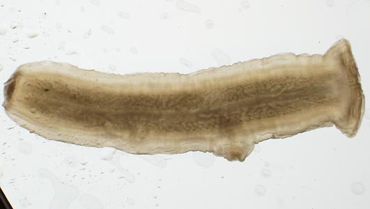
Figure 2: T. saginata proglottid revealing >13 primary uterine branches. www.cdc.gov
Figure 1: T. saginata proglottid revealing prominent genital pore. www.cdc.gov
Lifecycle
The lifecycle of T. saginata begins with the ingestion of undercooked beef containing the cysticerci larvae of Taenia saginata. Once the organisms has been ingested by the host, the cysticerci larvae develops in the intestines over several months. Once developed the adult worm attaches to the wall of the intestines and begins producing gravid (filled with eggs) proglottids. Those filled proglottids are then passed in the feces of the host where they can survive for days to months in the appropriate environment.
Viable proglottids are then ingested by cattle while grazing which release onchospheres from eggs and develop into cysticerci that encyst into the cattle’s muscle.
Morphology:
Taenia saginata is a tapeworm which belongs to the class cestoda, Cestodes are a type of flatworm which belong to the phylum Platyhelminthes.
Tapeworms are distinguished by their proglottids which give the worm a segmented appearance.
The egg of Taenia saginata measures approximately 40 microns in diameter. Once mature, the adult worm can reach up to 25 meters, but is typically measures 5 meters or less.
To distinguish T. saginata from T. solium (another common tapeworm), two distinct morphological features can be observed:
T. saginata has a 15-30 primary uterine branches, while T. solium has only 7-13 primary uterine branches.
The scolex of T. saginata is “unarmed,” meaning it has NO hooks. While T. solium is “armed,” meaning the scolex does have hooks.
Pathology:
Symptoms of T. saginata infection can range from a light or recent infection being asymptomatic, to a heavier infection being much more severe. Heavy T. saginata infection can cause abdominal pain, diarrhea, malnourishment, and weigh loss. Additionally, laboratory testing can reveal an elevated eosinophil.
Diagnosis:
To diagnose a T. saginata infection a specimen first needs to be properly collected using a Formalin / PVA collection kit for routine parasitology testing. Once a stool is collected using these two collection vials, the formalin preserved specimen can be concentrated using a flotation or sedimentation procedure to isolate any proglottids present.
Once the proglottid is isolated, the specimen can be visualized microscopically using a wet mount, or injected with a dye, such as India ink, to visualize the number of primary uterine branches.
In addition to microscopic identification of this organism, newer techniques have been developed to allow for the detection of specific DNA signatures. Isolating DNA from stool can be difficult as it can have lots of contaminants and interfering substances, but with a clean sample of parasitic nucleic acid, a rapid identification can be achieved with little operator training.
References:
www.cdc.gov
MLAB 1231 Lecture Notes – Presented by D. Scott Brewster


